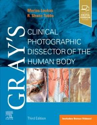
Gray's Clinical Photographic Dissector of the Human Body, 3rd Edition
Date of Publication: 11/2024
**Selected for 2025 Doody’s Core Titles® in Anatomy/Embryology**
The perfect hands-on reference, Gray's Clinical Photographic Dissector of the Human Body, 3rd Edition, is a practical resource in the anatomy lab, on surgical rotations, during clerkship and residency and beyond! This fully revised third edition uses a unique, step-by-step presentation of full-color cadaveric photographs to orient you more quickly in the anatomy lab, and points out the clinical relevance of each structure and every dissection. Each photograph depicts clearly labeled anatomical structures, including muscles, bones, nerves, blood vessels, and organs—making this one-of-a-kind resource ideal for preparing for laboratory sessions and as a useful reference during dissections.
The perfect hands-on reference, Gray's Clinical Photographic Dissector of the Human Body, 3rd Edition, is a practical resource in the anatomy lab, on surgical rotations, during clerkship and residency and beyond! This fully revised third edition uses a unique, step-by-step presentation of full-color cadaveric photographs to orient you more quickly in the anatomy lab, and points out the clinical relevance of each structure and every dissection. Each photograph depicts clearly labeled anatomical structures, including muscles, bones, nerves, blood vessels, and organs—making this one-of-a-kind resource ideal for preparing for laboratory sessions and as a useful reference during dissections.
Key Features
- Contains nearly 1,100 full-color photographs for comparison to the cadavers you study, helping you become more proficient and confident in your understanding of the intricacies of the human body
- Guides you through each dissection step-by-step, using a unique, real-world photographic presentation
- Includes complementary high-quality schematic drawings throughout to help orientate you and aid understanding
- Contains superb corresponding Gray’s illustrations to add clarity to key anatomical structures
- Helps you easily relate anatomical structures to clinical conditions and procedures
- Features new explanatory videos of human cadaveric dissection for each chapter
- Depicts the pertinent anatomy for more than 30 common clinical procedures such as prosthetic hip replacements, intravenous catheters, lumbar puncture, and knee joint aspiration, including where to make the relevant incisions
- Reflects the same level of accuracy and thoroughness that has made the Gray’s ‘family’ of products the most trusted learning resources in anatomy
- Prepared by an expert author team—highly experienced educators and leading authorities in clinical anatomy
- An eBook version is included with purchase. The eBook allows you to access all of the text and figures, with the ability to search, customize your content, make notes and highlights, and have content read aloud
The Evolve Instructor site with downloadable images is available to instructors through their Elsevier sales rep or via request at https://evolve.elsevier.com
Author Information
By Marios Loukas, MD, PhD, Dean School of Medicine, Professor, Department of Anatomical Sciences, Professor Department of Pathology, St. George's University, School of Medicine, Grenada, West Indies; Dean, College of Medical Sciences, Nicolaus Copernicus Superior School, Olsztyn, Poland and R. Shane Tubbs, PhD, MSc, PA-C, Professor, Director of Surgical Anatomy, Tulane University School of Medicine, Program Director of Anatomical Research, Clinical Neuroscience Research Center, Department of Neurosurgery, Department of Neurology, Tulane University School of Medicine, Department of Structural and Cellular Biology, Department of Surgery, Tulane University School of Medicine, USA


